
Figure 5.
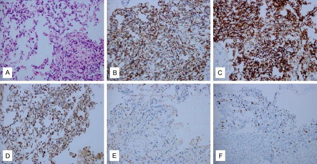
Histopathology of the angiosarcoma. A: Cellular area with atypical endothelial cells; B: Immunohistochemical stain for CD31 revealing positive endothelial cells; C: Immunohistochemical stain for CD34 revealing positive endothelial cells; D: Immunohistochemical stain for CK revealing positive; E: Immunohistochemical stain for factor VIII-related protein revealing positive endothelial cells; F: Ki-67 about 15%.