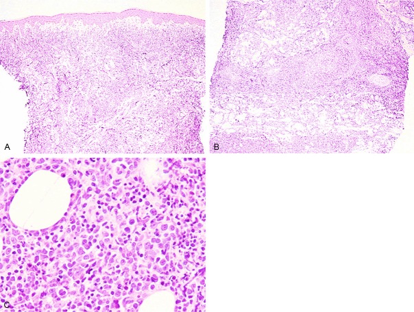
Figure 2.
Histopathological features of the knee nodule. A, B: Diffuse proliferation of lymphoid cells invading into the entire dermis and subcutis. No epidermal involvement is noted. HE, x 40. C: Large-sized atypical lymphoid cells with cleaved nuclei containing conspicuous nucleoli are present among medium-sized lymphoid cells with convoluted nuclei. HE, x 400.

