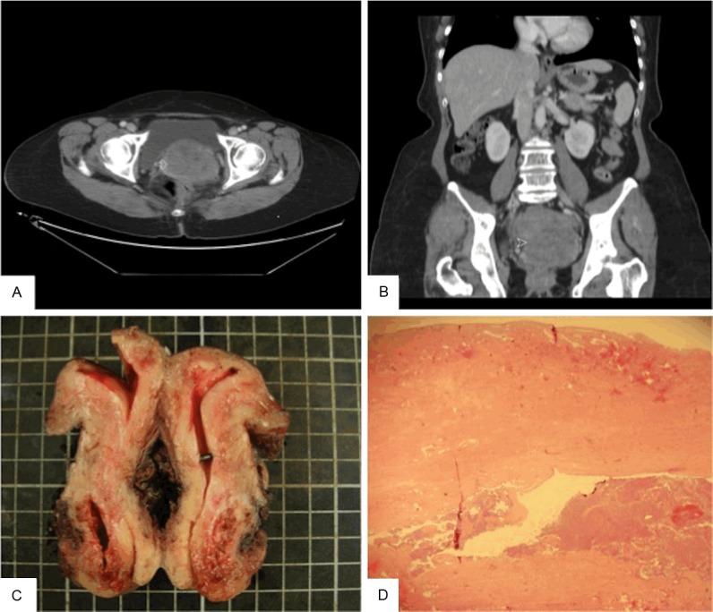
Figure 1.

A: The axial image post enhancement shows confluent uterine masses (arrow) with heterogeneous enhancement. The largest one measures 6.8 cm × 7.8 cm × 8.8 cm (depth × width × length in size). B: Coronal reformation of the above uterine masses (arrowhead). C: Grossly, the tumor is located on the posterior aspect of the uterine cervix while the endometrium and cervical canal are spared. D: The cervical mucosa is spared while the tumor rests at the underlying stroma.
