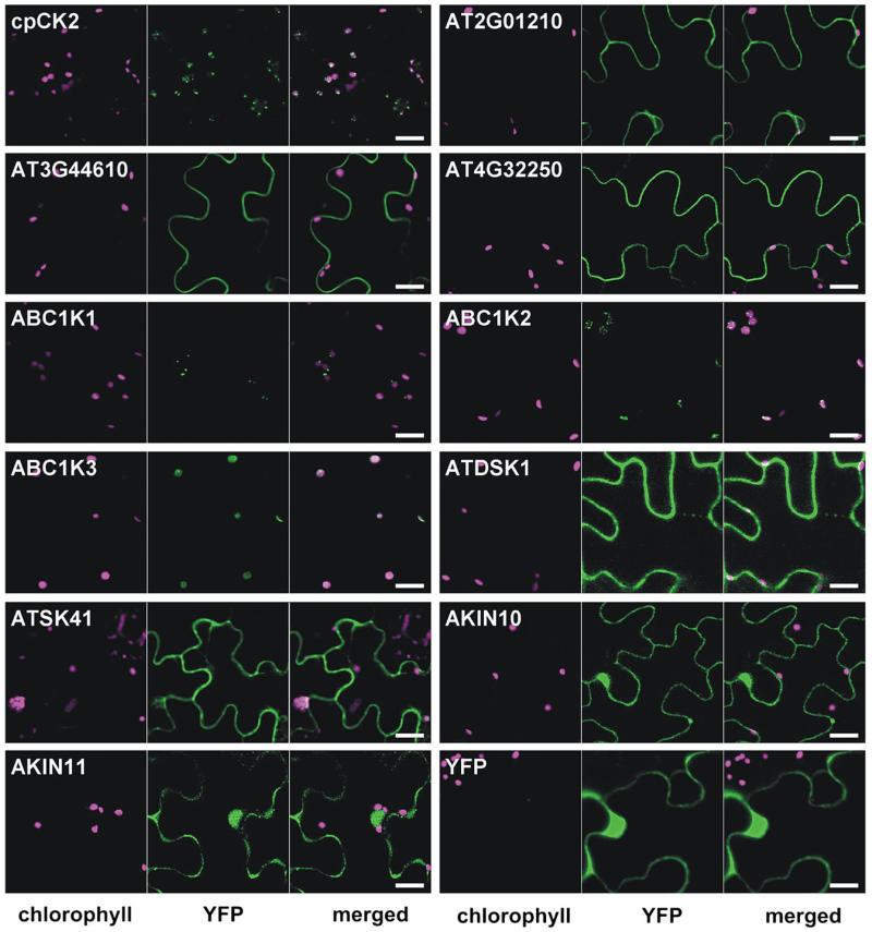
Fig. 1.
Localization analysis of selected protein kinases. Tobacco leaves infiltrated with genes of interest fused in front of YFP in the plant expression plasmid pBIN19 were analysed by confocal laser scanning microscopy 2 d after infiltration. Chlorophyll autofluorescence (magenta) is shown in the first channel and the YFP signal (green) in the second channel. The third channel shows the merged image. Scale bar=20 μm.

