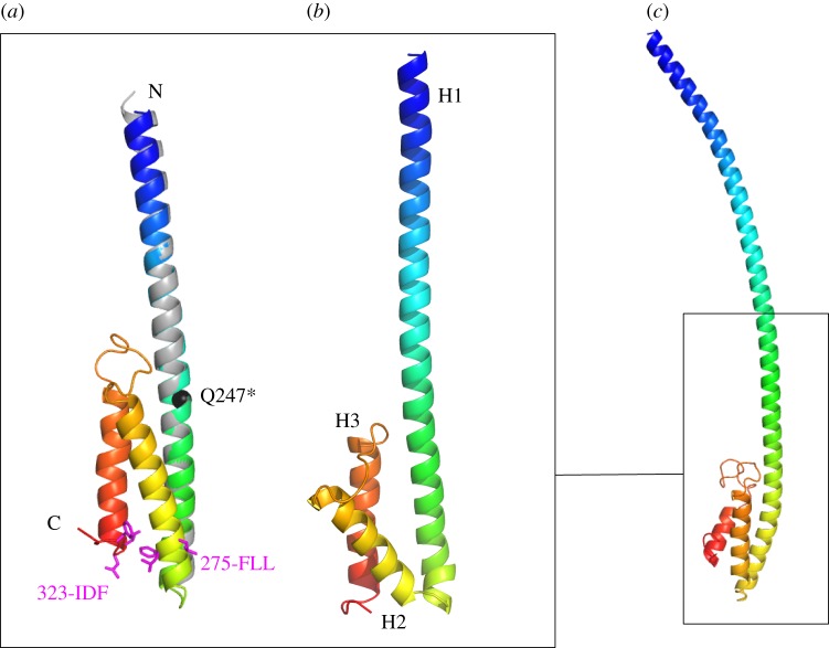
Figure 5.
Ab initio modelling of MuRF1 COS-box. (a) Top Quark model spanning MuRF1CC plus the subsequent COS-box region coloured in a blue-to-red gradient. The model is superimposed on the crystal structure of MuRF1CC (grey). The pathogenic Q247* mutation is shown in black and motifs previously identified to mediate microtubule binding in protein MID1 are in magenta [7]. Additional Quark models are shown in the electronic supplementary material, figure S2. (b) Rosetta-calculated model of the same segment derived from a cluster of 150 models. A comparison of Quark and Rosetta models is shown in the electronic supplementary material, figure S2. (c) Top Quark model of the full-length HD of MuRF1 (the degree of bending of the long helix H1 is not meaningful).

