Figure 6.
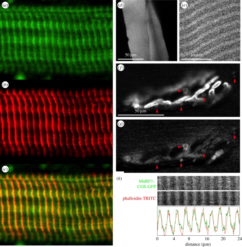
MuRF1 COS-box targeting of myocellular structures in vivo. (a–c) Localization of endogenous MuRF1 within sarcomeres. Muscle tissues were dissected from mice 5 days post-denervation, single myofibrils prepared from M. gastrocnemius and immunostained for (a) MuRF1 or (b) desmin. A merge of (a) and (b) indicates that endogenous MuRF1 under muscle stress targets predominantly (c) the Z-disc region and to a lesser extent the M-line. (d–g) In vivo targeting of transiently expressed MuRF1 COS-GFP fusion protein in transfected skeletal muscle. Depicted is (d,e) the enrichment of COS-GFP in sarcomeric striations as well as (f,g) an enrichment in puncta containing endocytic AChR ((f) α-bungarotoxin staining; (g) COS-GFP; arrowheads mark COS puncta that are also positive for AChR). A negative control corresponding to transfected GFP alone is shown in the electronic supplementary material, figure S6. (h) After fixation and staining with phalloidin-TRITC against f-actin, longitudinal sections of the muscles depicted in (d–g) show enrichment of COS-GFP in the Z-line/I-band, mimicking the distribution of endogenous MuRF1. Faint immunopositive signal on the M-line (a) is also present with COS-GFP in vivo (e) but does not stand fixation and staining (h).

