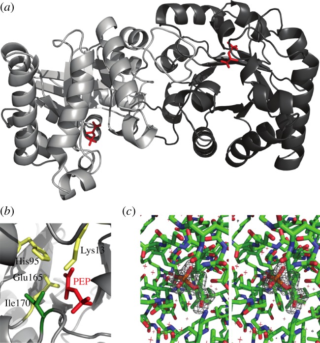
Figure 1.

Co-crystal structure of TPI with bound PEP. (a) Schematic of the TPI–PEP crystallographic structure. PEP locates in the active centre of both subunits in the asymmetric TPI dimer. (b) The catalytic pocket of TPI bound to PEP. Catalytic residues are highlighted in yellow, PEP in red, isoleucine 170 in green. (c) Stereoscopic illustration of the PEP binding site environment including a difference map in which PEP has been removed from the model and was refined against the experimental data for five cycles. The map has been contoured at 4 s.d. and reveals positive density for the missing ligand.
