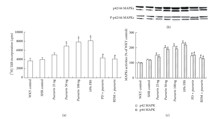
Figure 4.
Effect of puerarin on proliferation and activity of p42/44 mitogen-activated protein kinases (MAPKs) in microvascular endothelial cells (MECs). (a) MECs proliferation detected by [3H]-TdR incorporation. (b) Western blot analysis of phosphorylation of p42/44 MAPKs in MECs (upper panel). Equal protein loading was verified by reblotting with antitotal p42/44 MAPK antibody (lower panel). (c) Densitometry of the immunoblots shown in (a). Data are mean ± SD, *P < 0.05 compared with SHRs control, # P < 0.05 compared with puerarin treatment [100 ng], and n = 3.

