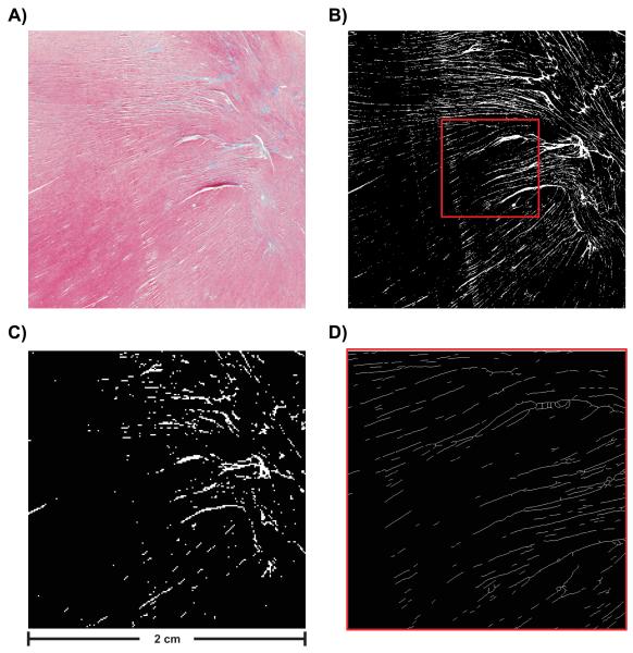
Fig. 3.
(A) Histological image selected for building FE meshes. Image consists of 2000 × 2000 pixels (pixel size 10 × 10 μm2). (B) Segmentation result for high resolution image. (C) Segmentation result for lower resolution image. Image consists of 200 × 200 pixels (pixel size 100 × 100 μm2). (D) Visualization of extracted skeleton showing a magnification of the area indicated by the red square in panel B).

