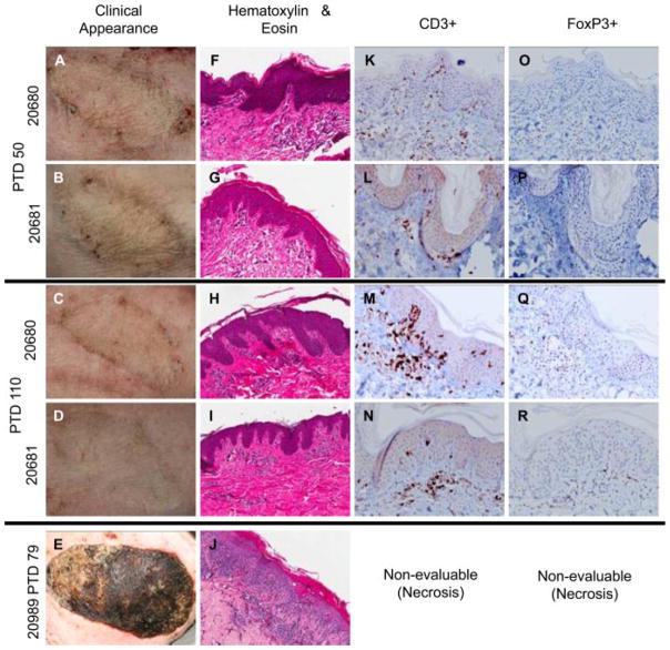
Figure 4. Induction of vascularized composite allograft tolerance achieved with contemporaneous HCT.
(A–D) Recipients of HCT and VCA displayed no clinical signs of VCA rejection at any time point, although Banff stage 1/2 rejection was diagnosed on biopsy at day 50 (F, G), this resolved by the subsequent biopsy following day 100 (H, I) and did not recur. In contrast, signs of rejection could be identified as early as day 14 in a VCA recipient which did not receive HCT (20989), while still under immunosuppression, progressing to complete rejection and necrosis by day 79 (E, J). Immunohistochemical analysis at day 50 revealed the presence of CD3+ infiltrates (K, L), which persisted beyond day 100 (M, N) but which were not associated with histological evidence of tissue damage. (O–R) FoxP3+ cells could be identified within these foci of CD3+ cells at all time points tested. Immunohistochemical staining of the rejected VCA in control animal 20989 was nonevaluable due to necrosis. HCT, hematopoietic cell transplantation; VCA, vascularized composite allograft.

