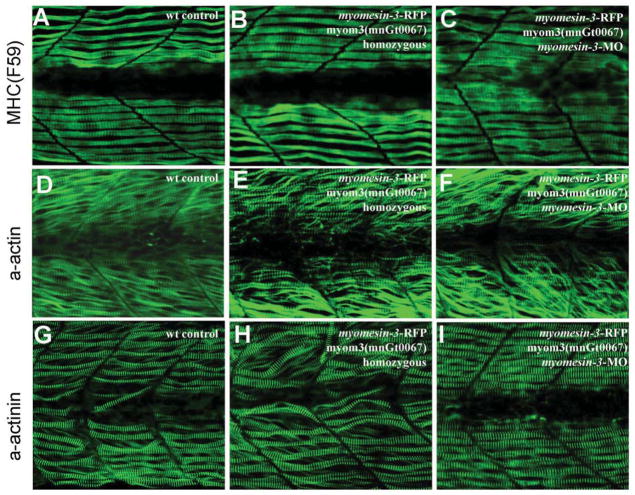
Fig. 8. The sarcomere organization in slow muscles of homozygous myomesin-3-RFP and myomesin-3 knockdown zebrafish embryos.
A–C: anti-MyHC antibody (F59) staining shows the thick filament organization in slow muscles of the control (A), homozygous myom3(mnGt0067) (B), or myomesin-3 knockdown (C) embryos at 28 hpf. D–F: α-actin immunostaining shows the organization of thin filaments in slow muscles of the control (D), homozygous myom3(mnGt0067) (E), or myomesin-3 knockdown (F) embryos at 28 hpf. G–I: α-actinin immunostaining shows the organization of Z-lines in slow muscles of the control (G), homozygous myomesin-3-RFP (H), or myomesin-3 knockdown (I) embryos at 28 hpf.

