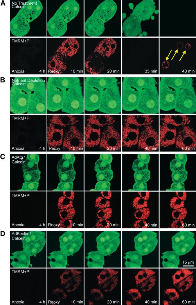
Fig. 3.
MPT inhibition by enhanced autophagy. After 4 hours of anoxia, hepatocytes were reoxygenated, and confocal images of calcein (green) and TMRM and PI (red) were simultaneously collected at 10, 20, 40, and 60 minutes. (A) Hepatocytes were reoxygenated with no other treatment. At the end of anoxia, calcein was excluded by mitochondria, and this was indicative of permeability pore closure. After 10 minutes of reoxygenation, mitochondria became repolarized without calcein redistribution into mitochondria. After 20 minutes, calcein redistributed into mitochondria, indicating onset of the MPT, and TMRM began to disappear, indicating mitochondrial depolarization. Within minutes, cytosolic calcein was lost, and PI labeled nuclei (arrows), indicating cell death. (B) Under the condition of nutrient depletion, mitochondrial voids in the calcein fluorescence were persistently present, and TMRM was continuously evident throughout reoxygenation. (C) AdAtg7 or (D) AdBeclin-1 infection prevented the MPT and cell death.

