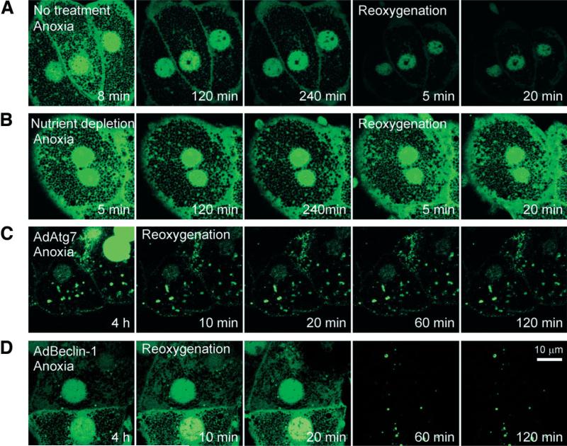
Fig. 4.
Autophagosome formation after A/R. Hepatocytes infected with AdGFP-LC3 were exposed to A/R, and changes in autophagosome formation were monitored by confocal microscopy. (A) After 8 minutes of anoxia, autophagosomes (green-fluorescing, punctate structures) were evident. After prolonged anoxia, most autophagosomes disappeared. Note a further decrease in autophagosomes after reoxygenation. (B) Nutrient depletion delayed loss of autophagosome. Autophagosomes were persistently present in hepatocytes coinfected with (C) AdAtg7 or (D) AdBeclin-1.

