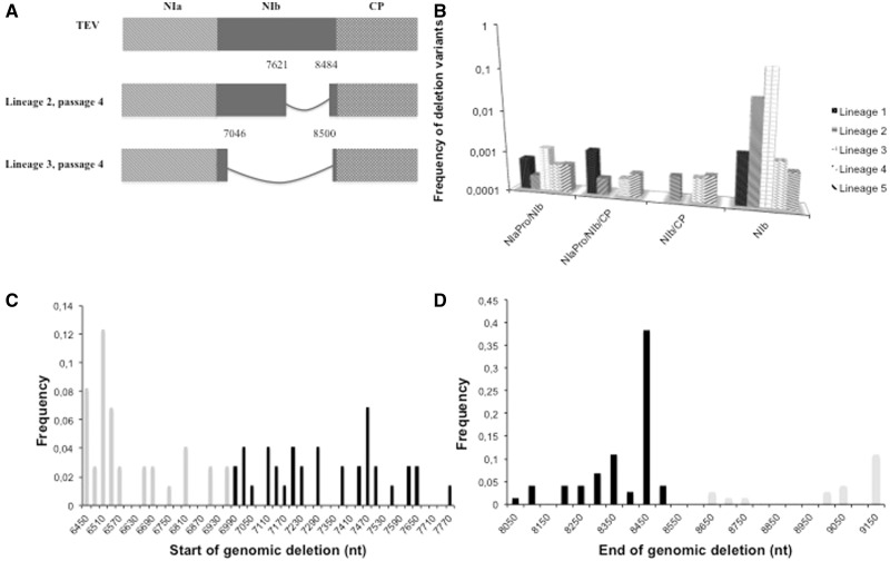
Fig. 2.—
(A) TEV and deletion variants detected by RT-PCR in different lineages at passages one and four. After four 3-week passages, NIb deletion variants are observed in different lineages as, for example, Lineage 4 and Lineage 5. All deletions observed after RT-PCR are situated within NIb and retain the reading frame. (B) Histogram of the frequency of different deletion variants observed in the five evolved lines. (C) Frequency distribution of the start nucleotide position of genomic deletions in the evolved lineages. (D) Frequency distribution of the end nucleotide position of genomic deletions in the evolved lineages. In (C) and (D), black bars indicate positions within the NIb cistron, gray bars indicate regions outside NIb, that is, in (C) within the NIaPro cistron and in (D) within the CP cistron.

