
Figure 3.
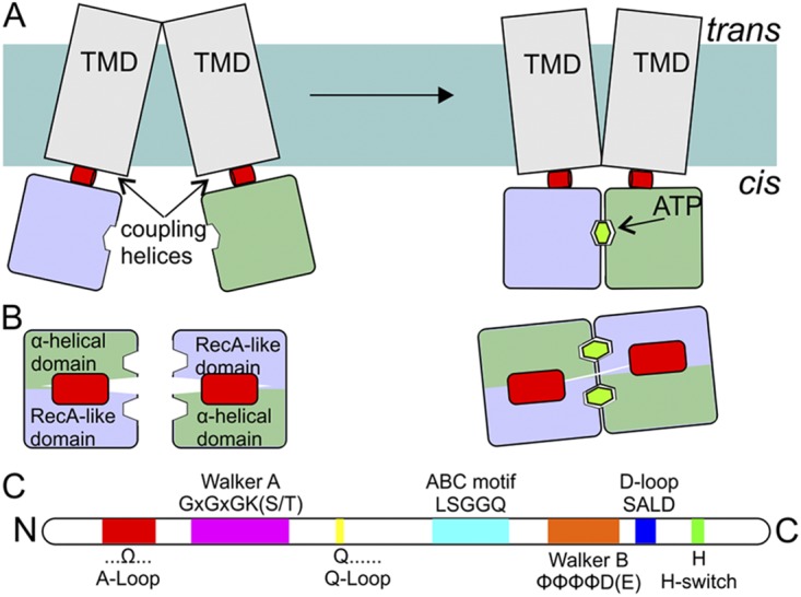
Schematic representation of NBDs and coupling helices. (A) Side view (from the membrane plane) of an ABC transporter. NBDs (blue and green; colors of the domains are as in Fig. 2) are attached to the TMDs (gray) via so-called coupling helices (red) present in loops of the TMDs. ATP binding and hydrolysis cause rearrangements in the NBDs, which are propagated to the TMDs via the coupling helices. (B) Top view (along an axis perpendicular to the membrane) of the NBDs and the coupling helices from the TMDs. (C) The relative positions of sequence motifs in NBDs (see also Fig. 2).