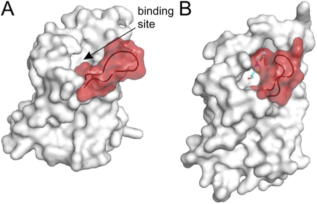
Figure 6.

Structural changes in the S-component of ECF transporters (substrate-binding TM protein of ECF transporters). The loop between TM helices 1 and 2 is highlighted in red. (A) S-component from the ECF–HmpT complex (Protein Data Bank accession no. 4HZU) in the substrate-free form. The shape of the binding pocket is visible, indicated by the arrow. (B) Thiamin-specific ThiT (Protein Data Bank accession no. 3RLB) in the substrate-bound state. The loop between TM helices 1 and 2 closes the binding pocket. The substrate thiamin is shown as sticks.
