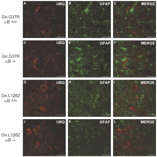
Figure 5.
Ubiquitin-positive structures do not change in location or abundance in the absence of αB-crystallin. Mice were perfused with 4% paraformaldehyde and spinal cord tissue was immersed in sucrose prior to cryostat sectioning. Sections were stained with ubiquitin (A, D, G, J) and GFAP (B, E, H, K), followed by staining with secondary fluorescent antibodies: anti-rabbit-AlexaFluor 568 (A, D, G, J) and anti-mouse-AlexaFluor 488 (B, E, H, K). Ventral horn of spinal cord is shown at an original magnification of 40x. The image shown is representative of at least 3 repetitions of the experiment. Scale bar – 40 microns.

