Figure 3.
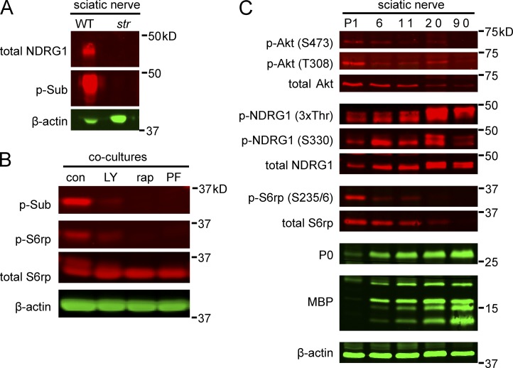
S6rp and NDRG1 correspond to the 30- and 45-kD phospho-substrate bands. (A) Western blots of wild-type and stretcher sciatic nerve lysates were probed with anti-NDRG1 and p-Sub antibodies; β-actin serves as a loading control. NDRG1 and the p45-kD p-Sub doublet are not detected in the stretcher nerve lysates. (B) Western blots of lysates from nonmyelinating co-cultures were treated with 50 nM rapamycin, 20 µM LY294002, or 40 µM PF-4708671 for 18 h and then probed with p-Sub, p-S6rp, and total S6rp antibodies. p-S6rp and p30 are not detectable in co-cultures treated with rapamycin, LY294002, or PF-4708671. (C) Expression of phospho- and total Akt, NDRG1, and S6rp in developing rat sciatic nerves. Lysates of postnatal sciatic nerves were probed on Western blots with the indicated antibodies. Both total and phosphorylated NDRG1 are up-regulated in parallel with compact myelin proteins P0 and MBP. S6rp and Akt are maximally expressed and phosphorylated early, then down-regulated as myelination proceeds.

