Figure 4.
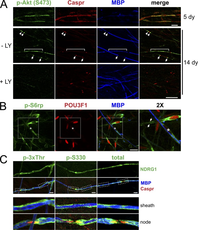
Localization of p-S6rp, p-NDRG1, and p-Akt in myelinating co-cultures. (A) Co-cultures maintained in myelinating media for 5 or 14 d were stained for p-Akt (green), Caspr (red), and MBP (blue). In 5-d co-cultures (top row), Schwann cells just before myelinating express p-Akt (S473) along their length; Caspr staining of axons is diffuse and MBP is not yet expressed. In 14-d co-cultures (bottom two rows), p-Akt staining varied depending on the maturity of the myelin segments. In a thinly myelinated segment with a single paranode (bracket), p-Akt was prominent along the Schwann cells, in more mature segments with two hemi-paranodes (arrows) or surrounding a mature node (arrowheads) staining was concentrated in the paranodal and juxtaparanodal regions. Addition of the LY inhibitor before fixation abolished p-Akt staining. Bar, 20 µm. (B) Myelinating co-cultures were stained for p-S6rp (green), POU3F1 (red), and MBP (blue). p-S6rp was maximally expressed in the cytoplasm and along the processes of a premyelinating Schwann cell expressing POU3F1 (arrows) and at lower levels in myelinated Schwann cells (one is marked by an asterisk). Bar, 20 µm. Inset (far right panel) is at 2×. (C) Myelinating co-cultures stained for phospho- (3xThr, S330) and total NDRG1 (all green), MBP (blue), and Caspr (red). Myelinating Schwann cells express phospho- and total NDRG1 (green) along the outside of the myelin sheath and in some paranodes (insets, 3× magnification). Bar, 20 µm.

