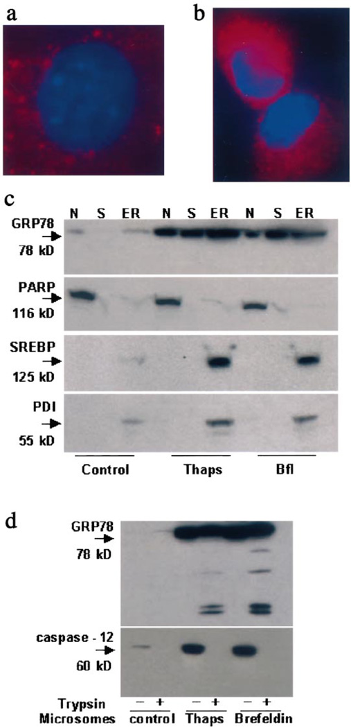
Fig. 3.
Expression and localization of GRP78 before and after ER stress. Double Immunofluorescent staining for GRP78 before (a) and after ER stress (b) with Cy3 in 293T cells left in control medium for 12 h before fixation in buffered 4% paraformaldehyde. For the detection of GRP78, cells were stained with rabbit polyclonal anti-GRP78 antibody as primary antibody and Cy3-labeled donkey anti-rabbit IgG as the secondary antibody. Nuclei are counterstained using VectaShield® Mounting Medium with DAPI (Vector Laboratories, Burlingame, CA, USA). c: Western blots of nuclear (N), cytoplasmic (S) and microsomal (ER) fractions of 293T human renal epithelial cells before and after treatment with 2.5 µM brefeldin-A or 2.5 µM thapsigargin for 24 h. Cellular fractions were also probed for standard marker proteins namely PARP (nuclei), PDI and SREBP-1 (ER). d: Tryptic cleavage of GRP78 in isolated microsomes. Microsomes isolated from control, thapsigargin- (thaps) and brefeldin-A-treated cell extracts were digested by 0.05% trypsin-EDTA for 30 min at 35°C. At the end of the reaction, samples were detected by Western blotting.

