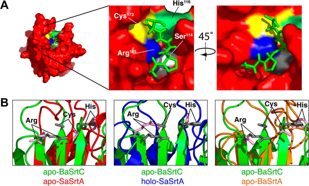
Figure 6.
(A) Model of the CWSS peptide structure from the SaSrtA–LPAT* complex on the substrate binding surface of BaSrtC. The peptide (green) is taken from the SaSrtA–LPAT* structure without modification. Active site residues are indicated along with the potential Ser114 binding site. The hydroxyl oxygen of Ser114 (Ser114 is colored gray, while its OH group is colored white) comes into close contact with the side chain of position X. (B) Close-up overlay of the BaSrtC structure (green) with apo-SaSrtA (red, left), holo-SaSrtA (blue, center), and apo-BaSrtA (orange, right). Active site arginine, cysteine, and histidine residues are denoted. The active site residues of apo-BaSrtC overlay best with those of holo-SaSrtA (SaSrtA–LPAT*).

