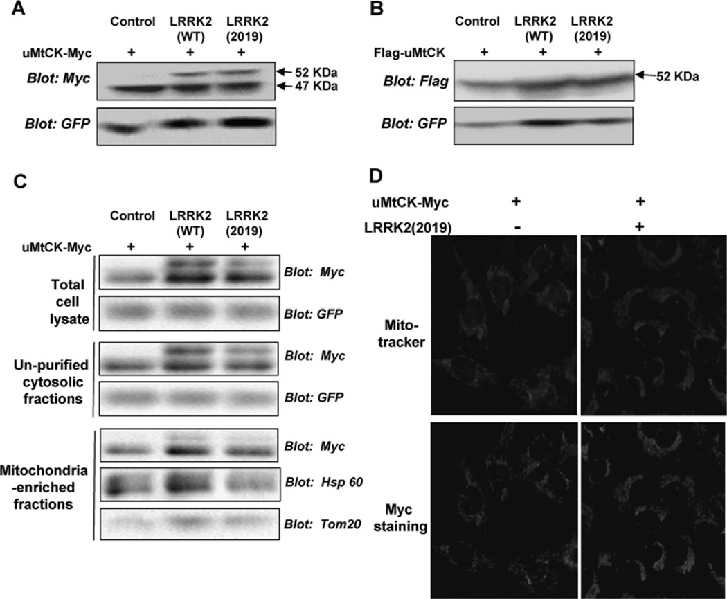
Figure 3. LRRK2 expression leads to the accumulation of an unprocessed immature form of uMtCK.
(A) pcDNA3-uMtCK-Myc was transfected either alone or together with LRRK2 WT or LRRK2 2019S into HEK-293 cells. At 48 h later, cell lysates were analysed with an anti-Myc antibody. Expression of both LRRK2 WT and LRRK2 2019 leads to the accumulation of a higher-molecular-mass form of uMtCK-Myc. (B) pCMV-Flag-uMtCK was transfected alone or together with LRRK2 WT or LRRK2 2019S into HEK-293 cells. Forty-eight hours later, cell lysates were analysed using an anti-Flag antibody. Only the 52 kDa Flag-uMtCK bands were observed. (C) HEK-293 cells were transfected with pcDNA3-uMtCK-Myc either alone or together with LRRK2 WT or LRRK2 2019S. Forty-eight hours later, differential centrifugation was performed for the cell lysates. The total cell lysates, crude cytosolic fractions and mitochondria-enriched fractions were analysed with antibodies specific for Myc, GFP, Hsp60 and Tom20. (D) U2OS cells were transfected as in (A). MitoTracker Red dye was used to detect mitochondria. Location of uMtCK-Myc was shown by immunostaining with Myc antibody.

