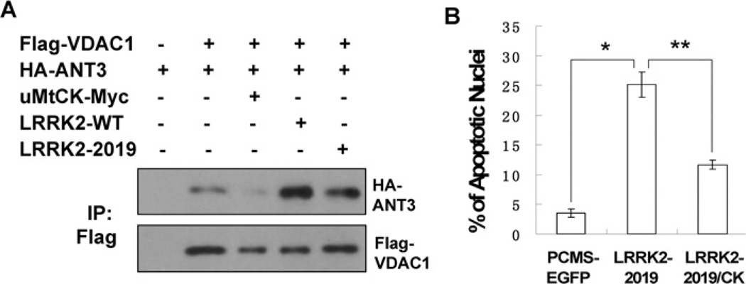
Figure 4. LRRK2 promotes the interaction between ANT and VDAC and uMtCK can suppress LRRK2-induced apoptosis.
(A) Flag-VDAC1, HA-ANT3, uMtCK-Myc, LRRK2-WT and LRRK2-2019S were transected into HEK-293 cells either alone or in combination as indicated. Thirty-six hours later, the cell lysates were immunoprecipatated with an anti-Flag antibody. The immunocomplexes were analysed with anti-HA and anti-Flag antibodies. (B) pCMS-EGFP, pCMS-EGFP-LRRK2-2019 was transfected either alone or together with uMtCK-Myc into neuronally differentiated PC12 cells. Forty-eight hours later, the cell nuclei were stained with Hoechst 33342. The percentage of transfected cells exhibiting apoptotic morphology was calculated. Values represent means ± S.E.M. for three replicate cultures. *P<0.008 and **P<0.01.

