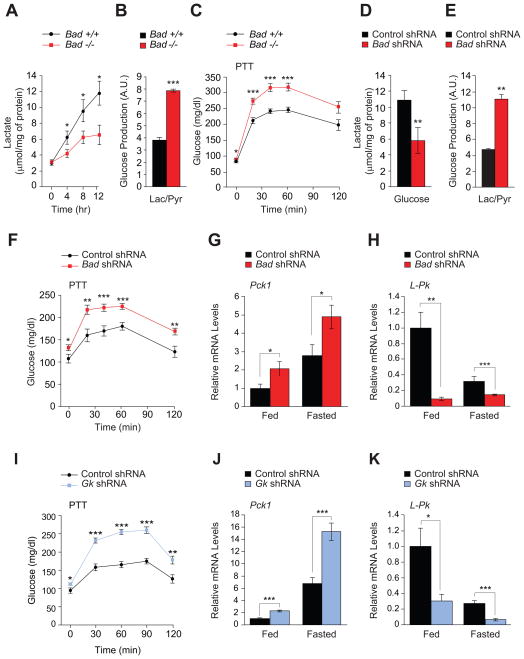
Figure 1. Hepatic glucose metabolism in the absence of BAD.
(A) Glucose-stimulated lactate production by primary Bad +/+ and −/− hepatocytes (n=5–8).
(B) Glucose release by Bad +/+ and −/− hepatocytes treated with lactate/pyruvate (n=6).
(C) PTT in Bad +/+ and −/− mice (n=14–20).
(D) Lactate production in Bad knockdown hepatocytes 8 hr after glucose stimulation (n=9).
(E) Glucose production in Bad knockdown hepatocytes treated with lactate/pyruvate (n=3–5).
(F) PTT in C57BL/6J mice after hepatic knockdown of Bad (n=12–17).
(G–H) Relative hepatic mRNA levels of Pck1 (G) and L-Pk (H) in fed and overnight fasted C57BL/6J mice after hepatic knockdown of Bad (n=10).
(I) PTT in C57BL/6J mice after hepatic knockdown of Gk (n=8–12).
(J–K) Relative hepatic mRNA levels of Pck1 (J) and L-Pk (K) in fed and overnight fasted C57BL/6J mice after hepatic knockdown of Gk (n=5).
Error bars, ± SEM. *p < 0.05; **p < 0.01, ***p < 0.001.
See also Figure S1.

