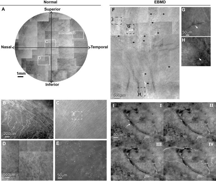
Figure 2.
A) Whole mount view of epithelial nerve distribution in the normal left cornea. Images were acquired in a time-lapse mode with a Nikon Eclipse TE200 and with a 5X lens in compliance with the natural shape of the cornea. The epithelial nerve bundles ran in a radial pattern from the periphery to merge in an area within the inferior-nasal quadrant beyond the corneal apex. Highlighted images showed the detailed architecture of the epithelial nerve distribution in the peripheral (B), central (C), and vortex areas (D). E) High magnification image recorded from the central area as marked in Figure 2C shows the nerve terminals at the superficial epithelia. F) Whole mount view of corneal epithelial nerve distribution in the central area of the inferior quadrant of the right cornea. The total area is about 45 mm2, consisting of 38 images recorded in a time-lapse mode with a 10X lens. Arrows indicate the pathological loci. G) and H) are highlighted images showing the detailed architecture of the epithelial nerve distribution in the central and vortex area as marked in F). I) Topographic images, recorded at the plane of superficial epithelia (i), wing cells (ii), basal cells (iii) and the basement membrane (iv), show detailed epithelial innervation in a finger-print lesion (arrow) at the central cornea.

