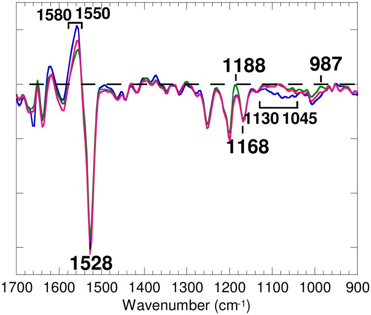
Figure 1.
The chromophore region (1700-900 cm-1) of the M-minus-BR spectrum (d2) of wild type at pH 7 (37, blue line), the spectrum of d2 of wild type at pH 5 (green line), and the calculated spectrum of the M-minus-BR component of wild type at pH 5 (red line) obtained by subtracting 40% of the spectrum of d0 (mainly due to the L-minus-BR spectrum) from the spectrum of d2. The M-minus-BR spectra at pH 7 and pH 5 were normalized on the basis of the negative band at 1168 cm-1. One division of the ordinate corresponds to 0.002 absorbance unit for bacteriorhodopsin at pH 7. The level of zero absorbance is shown by a horizontal broken line.

