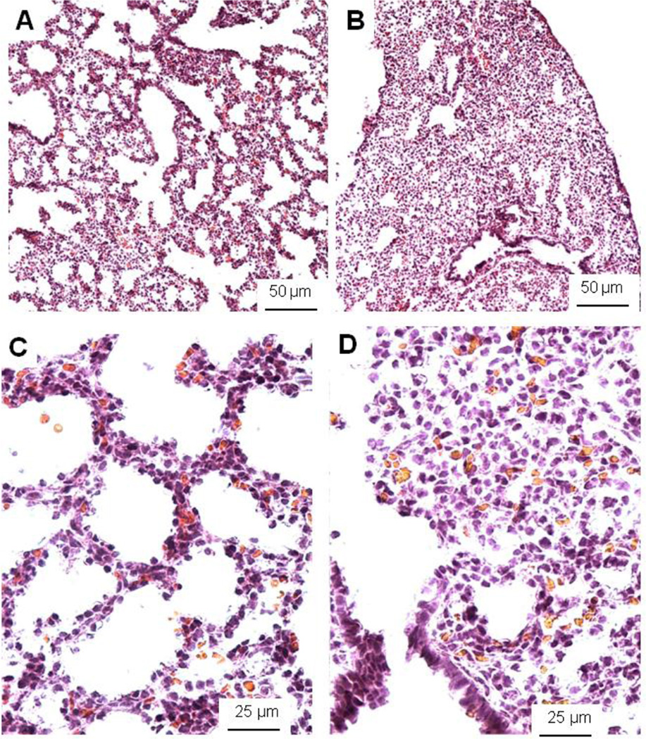
Figure 2.
Histological pictures of E17 control HER4heart+/− (A, C) and HER4heart−/− (B, D) lungs. Representative light microscopic images from cryosections stained with hematoxilin and eosin. HER4heart−/− (B) lungs have fewer transitory ducts and saccules compared to HER4heart+/− control lungs (A). A higher resolution shows more epithelial cells surrounding the air spaces in control lungs (C). HER4heart−/− (D) lungs have less sacculi and show accumulation of tissue containing mesenchyme cells. Morphometric evaluations revealed an increased volume density of the septal tissue and a decreased volume density of the airspace in HER4heart−/− lungs.

