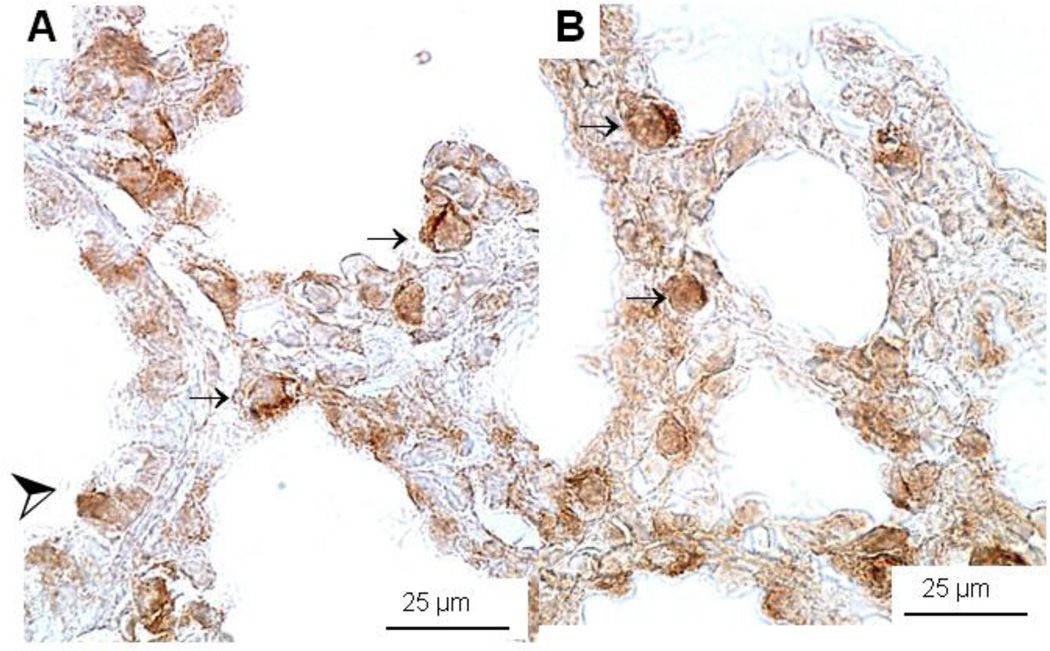
Figure 4.
Immunohistochemistry of SP-B protein expression in E18 control HER4heart+/− (A) and HER4heart−/− (B) fetal lung. Representative images of lung sections from HER4heart−/− lungs (B) compared with HER4heart+/− control lungs (A). Dark brown staining is seen in SP-B-positive cells which include type II epithelial (small arrows) and Clara cells (large arrow head).

