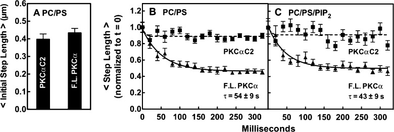
Figure 5.
Single-particle step lengths for freely diffusing full-length PKCα and the C2 domain bound to pre-DAG bilayers. Step sizes were quantitated by TIRF for single-particle tracks on supported bilayers of the indicated lipid composition. (A) Comparison of the average initial step lengths of PKCα and the C2 domain on PC/PS bilayers. The initial step length is the distance traveled per 20 ms frame at the beginning of a single-particle diffusion track (the second frame of the track is quantitated because the first frame often captures a partial step following particle binding), as determined by averaging the initial steps of at least 200 particle tracks (≥30000 steps) per experiment in three separate experiments (n ≥ 600). Data were normalized to the initial step length of FL PKCα (0.40 ± 0.04 μm). (B and C) Time dependence of step length for successive steps during the first 320 ms after the particle had bound to the indicated bilayer. With the second 20 ms step of the diffusion track as a starting point (see the legend of panel A), the length of the indicated step was determined for each diffusion track, and the lengths of corresponding steps were averaged over at least 200 particle tracks in three separate experiments (n ≥ 600) to yield the indicated time courses. Finally, the two time courses in each box were normalized to the initial step length of FL PKC. In all experiments, the free Ca2+ concentration was 6 μM in a physiological buffer (Materials and Methods) at 22 ± 0.5 °C.

