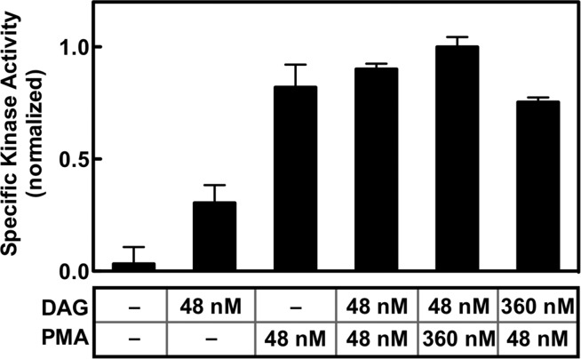
Figure 8.
Comparison of the effects of DAG and PMA on the PKCα kinase specific activity. The total PKCα kinase activity and the bound density of PKCα on the bilayer were measured as described in the legend of Figure 3 for membranes containing 3:1 PC/PS bilayers and the indicated levels of activating diacylglycerol and/or phorbol ester PMA (an activating lipid concentration of 48 or 360 nM corresponded to 2.0 or 7.5 mol % in the bilayer, respectively). Specific activity was calculated as the ratio of kinase activity to membrane-bound enzyme density under each bilayer condition. Each bar was calculated from two duplicates for each kinase and binding assay (n = 4). In all experiments, the free Ca2+ concentration was 6 μM in kinase assay buffer (Materials and Methods) at 22 ± 0.5 °C.

