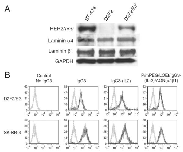
Fig. 2.
Expression level of anti-HER2/neu and the specific binding to human anti-HER2/neu. A) The expression of anti-HER2/neu, laminin α4, and β1 chains in BT-474, D2F2, and D2F2/E2 cell lines was detected by western blot analysis. Expression of GAPDH was used as a loading control. B) Binding of various molecules to D2F2/E2 (top) and SK-BR-3 (bottom) as determined by flow cytometry. Cell lines were incubated in control buffer (gray line) or in the presence of anti-HER2/neu IgG3, anti-HER2/neu IgG3-(IL-2), or P/mPEG/LOEt/IgG3-(IL-2)/AON(α4β1) (black line) all equivalent to 2 μg of IgG3, followed by rabbit anti-human κ conjugated to FITC.

