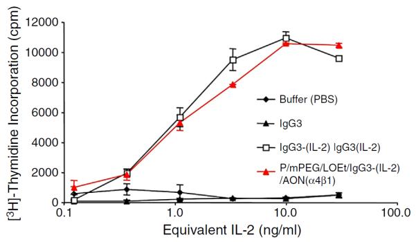
Fig. 3.

In vitro biologic activity of IL-2 within the PMLA-fusion nanobioconjugate. IL-2 activity was determined by incorporation of [3H]-thymidine into CTLL-2 cells incubated in the presence of varying concentrations of IL-2 fusion proteins and controls. Serial 1:3 dilutions of IL-2 equivalents ranging from 30 ng/ml to 0.13 ng/ml of anti-HER2/neu IgG3 (IgG3), anti-HER2/neu IgG3-(IL-2) [IgG3-(IL-2)], and PMLA-fusion nanobioconjugate P/mPEG/LOEt/IgG3-(IL-2)/AON(α4β1) were made in quadruplicate and incubated with CTLL-2 cells for 18 h followed by a 6-hour pulse of [3H]-thymidine. The error bars indicate the standard deviation.
