Figure 5.

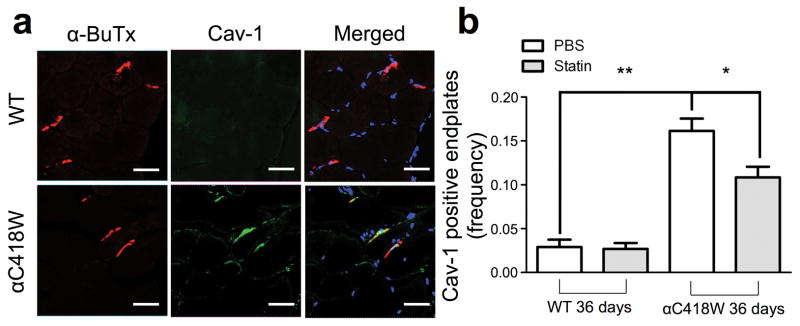
αC418W mice display a higher percentage of Cav-1 positive end-plates, which is in turn reduced by statin treatment. Tibialis anterior muscle cryo-sections were obtained from αC418W and WT mice. These were stained for nAChRs, Cav-1 and nuclei with α-bungarotoxin, immunohistochemistry using anti-Cav-1 antibodies and DAPI, respectively. (a) αC418W mice showed a significant amount of Cav-1 colocalizing with end-plates, as identified by nAChR staining, whereas WT mice displayed a significant lower amount (**P<0.01, n=3 for αC418W and WT mice on PBS treatment) (scale bars represent 36μm). (b) After 36 days of treatment, αC418W mice showed a significant reduction in the percentage of Cav-1-positive end-plates, whereas the WT percentages remained unchanged (*P<0.05, n=3 for αC418W and WT mice on PBS and statin treatment).
