
Figure 7.
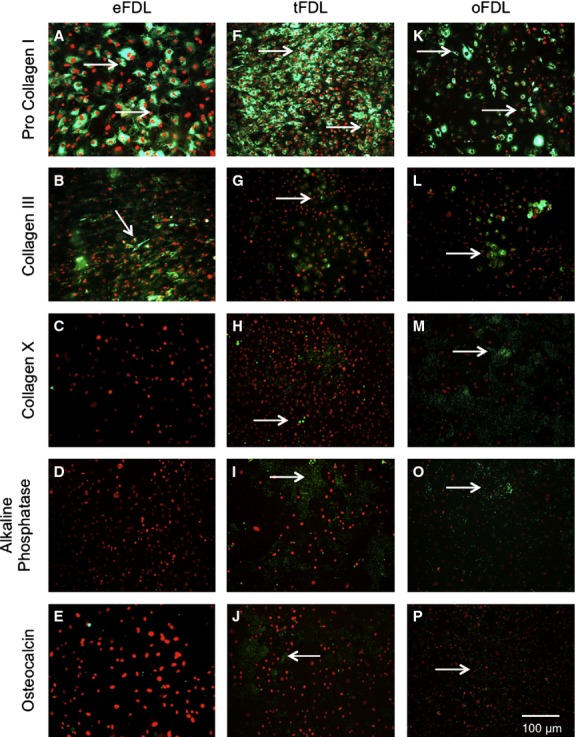
Immunological profile of cells from adult non-ossified FDL, adult ossified FDL and embryonic FDL. (A-E) Expression profile of embryonic tendon cells: a high expression of procollagen I (A) and collagen III (B), both part of the tendon extracellular matrix and no expression of collagen X (C), alkaline phosphatase (D) and osteocalcin (E), all markers for endochondral ossification and bone. (F-J) Expression profile of the non-ossified adult tendon cells: the expression of tendon markers is noticeably lowered (F,G), whereas bone and ossification markers have a slightly increased expression (H-J). (K-P) Expression profile of the ossified adult tendon cells: the expression of tendon markers is considerably lowered (K,L) and bone and ossification markers have a substantially strong expression (M-P).