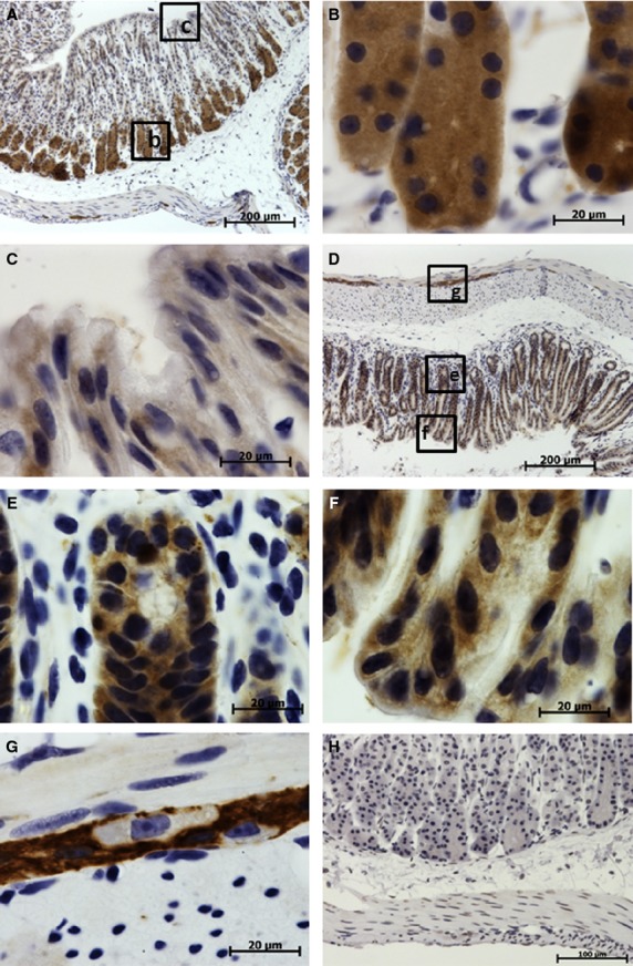
Figure 5.

Immunohistochemical studies (IHC) localization of translationally controlled tumor protein (TCTP) in the glandular portion of the stomach. Translationally controlled tumor protein expression is shown in brown, cell nuclei stained blue with hematoxylin. (A-G) Sections treated with anti-TCTP antibody. (H) Control section treated with irrelevant rabbit polyclonal antibody. (A) Low-power image of stomach fundus demonstrating high TCTP expression in the base of gastric glands and low expression in the neck and surface lining cells. (B) High-power image of the base of fundal glands with high TCTP expression. (C) High-power image of necks of fundal glands with low TCTP expression. (D) Low-power image of pyloric portion of the stomach. (E) High-power image of the base of pyloric gland with moderate to high TCTP expression. (F) High-power image of necks of pyloric gland and gastric pits demonstrating moderate TCTP expression. (G) High-power image of the myenteric nerve ganglion in the stomach muscular layer with high TCTP expression, note the absence of TCTP in the perikaryon of neurons.
