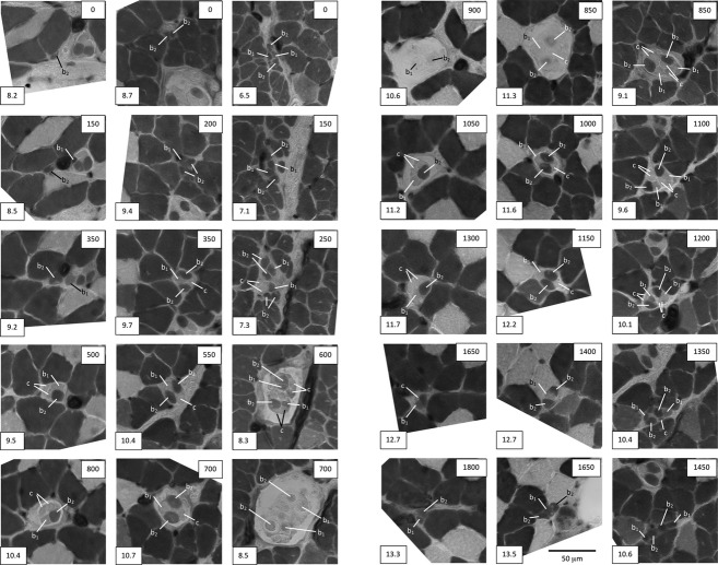
Figure 3.
Representative cross-sections taken at intervals throughout three sample spindles from a BEL soleus muscle to show the identification of intrafusal-fibre types, and to illustrate: (left column) the most common complement of one bag2, one bag1 and two chain fibres; (middle column) a spindle with two bag2, one bag1 and one chain fibre; and (right column) one of the largest spindles found in any strain with a complement of two bag2, two bag1 and four chain fibres. The box at the lower left of each micrograph gives the reference for the section in the format: slide number.section number. The box at the upper right gives the nominal distance from the first section in μm. In the spindle shown in the right column, the two bag2 fibres are thought to have branched somewhere between sections 7.1 and 7.3, resulting in the appearance of four profiles in 7.3. Acid-stable ATPase following pre-incubation at pH 4.47; differential staining properties of the three types of intrafusal fibre are described in the text.

