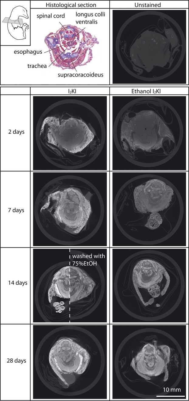
Figure 5.

MicroCT images of quail embryos stained with 3.75% I2KI and ethanol I2KI at various stain durations and a Mallory Trichrome histological section for comparison. All CT images are unmodified images obtained from tomographic reconstruction software and at the same scale. An embryo stained with I2KI for 14 days and subsequently washed with 75% ethanol is presented in the right side of an embryo stained with I2KI for 14 days for direct comparison. All the microCT images are from approximately the same cross-sectional level illustrated in the top left. The histological image is magnified 1.8 times to be comparable to a size of all microCT images. The extreme shrinkage of the histological sample is an artifact of dehydration and paraffin processing.
