
Figure 2.
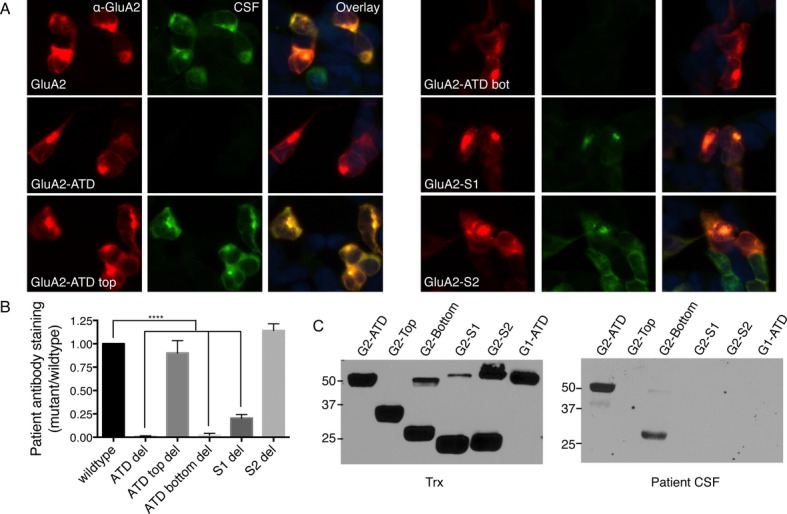
Antibodies from patients with anti-AMPAR encephalitis primarily recognize the bottom lobe of the ATD of GluA1 or GluA2. (A) Staining pattern of CSF from a typical anti-AMPAR encephalitis patient on HEK293 cells transfected with GluA2 deletion mutants shows a loss of reactivity with deletion of the bottom lobe of the ATD. (B) Quantification of deletion mutant CSF staining over four patients, three with primary GluA2 reactivity and one with primary GluA1 reactivity, using GluA2 or GluA1 mutants, respectively. (C) Reactivity with receptor domains on western blot. Left, fusion protein expression measured by anti-Thioredoxin antibody (Trx). Right, patient CSF reacts primarily with the ATD and the bottom lobe of the ATD. S1 deletion also decreases antibody binding, but does not appear to bind on western blot, possibly due to the proximity of the S1 domain to the bottom lobe of the ATD. *P < 0.05, ***P < 0.001, one-way ANOVA plus Dunnett's post hoc testing.