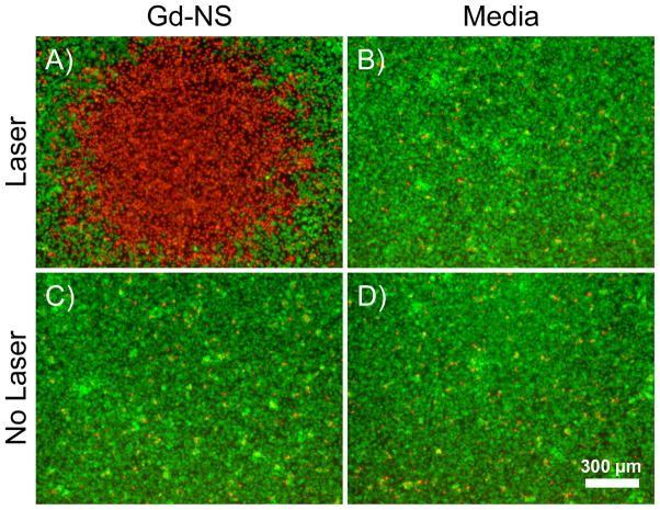
Figure 6.
(A) Gadolinium-nanoshells (Gd-NS) effectively ablated B16-F10 melanoma cells after particle incubation and NIR exposure (808 nm, 35 W/cm2, 3 min). Fluorescent viability staining was performed with calcein AM, which shows live cells in green, and ethidium homodimer-1, which depicts dead cells in red. The red area of cell death indicates the irradiation zone. (B) Cells irradiated under the same conditions with no prior particle incubation remained viable. Non-irradiated cells incubated (C) with and (D) without particles also remained viable. Scale bar = 300 μm.

