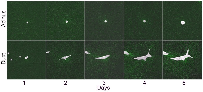
Figure 5. Time-course of epithelial morphogenesis.
Still images at a single position taken at day 1 (3 hours after seeding), 2 (27 hrs), 3 (51 hrs), 4 (75 hrs) and 5 (99 hrs) showing the formation of an acinus in 50% Matrigel (top row) and a duct in 0% Matrigel (bottom row). Brightfield and RCM images showing the collagen fibers are overlaid. Scale bar, 50 μm.

