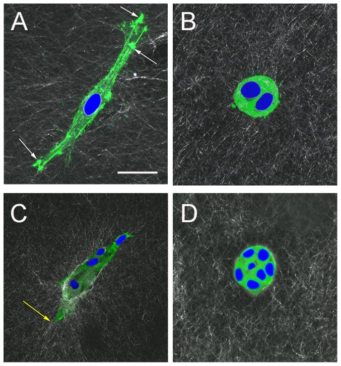
Figure 7. F-actin organization in epithelial cells forming acinar and ductal structures.
(A) and (B) are images at day-3 and (C) and (D) at day-5. Nuclei are stained with DAPI. (A) Elongated cell in 0% Matrigel. The presence of localized areas of actin particularly at the leading edges indicates actin-associated adhesion with the ECM (white arrows). (B) Rounded cells in 50% Matrigel. Few filopodia are observed as the majority of actin-staining originates from the cell cortex. (C) Duct in 0% Matrigel. Co-alignment between actin and extracellular collagen fibers is evident (yellow arrow). (D) Acinus in 50% Matrigel, as in (B) the majority of staining originates from the cell cortex and there are no filopodia. Scale bar, 25 μm.

