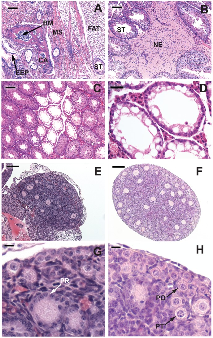
Figure 1. Histology of testes and ovaries from 129 mice exposed to cyclophosphamide at 7.5 mg/kg/day on E10.5 and 11.5.
(A-D) Testes from 4-week-old mice. (A) TGCT characterized as a teratoma originating from multiple dermal layers; (B) TGCT containing only neuroepithelial cells. Abbreviations: BM: bone marrow; CA: cartilage; EEP: endodermal epithelium; NE: neuroepithelial cells; MS: muscle; ST: seminiferous tubule. (C) non-TGCT-bearing testis showing active and atrophic tubules. (D) High magnification of atrophic tubules containing only Sertoli cells. (E-H) Ovaries from 7-day-old mice. (E, G) From a mouse treated on E10.5 and 11.5 with 7.5 mg CP/kg/day. (F, H) Control ovary of the same age. G and H are the magnified views from portions of E and F respectively, showing primordial (PO), primary-transitional (PT) and primary (PR) follicles. The bar represents 100 μm in A, B, C, E & F; 30 μm in D; 10 μm in G and H.

