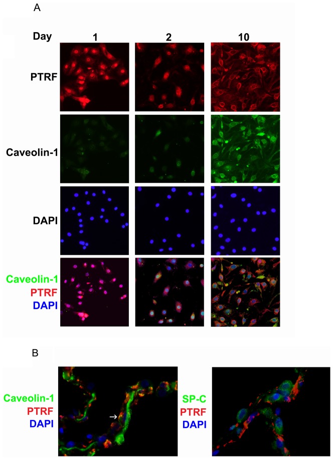
Figure 5. Sub-cellular localization of PTRF/CAVIN-1 changes from nuclear to cytoplasmic in transitioning hAT2 cells.
(A) Cells from two separate isolations were seeded on collagen-coated glass chamber slides and fixed on days 1, 2, and 10. PTRF/CAVIN-1 (red) and caveolin-1 (green) were detected by immunofluorescence; DAPI (nuclei, blue) was supplied in the mounting medium. Nuclear PTRF (day 1) becomes cytoplasmic and co-localizes with caveolin-1 by day 10. Results shown are representative. Original magnification 40X. (B) Paraffin-embedded normal human lung tissue sections were deparaffinized and immunofluorescence was performed for PTRF/CAVIN-1 (red) and caveolin-1 (green, left panel) or SP-C (green, right panel); DAPI (nuclei, blue) was supplied in the mounting medium. Caveolin and PTRF co-localize while PTRF and SP-C do not. Results shown are representative of three separate cellular preparations. Original magnification 100X.

