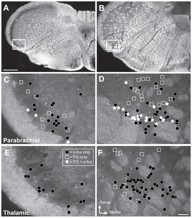
Figure 3.

Projection neurons are interspersed with neurons activated by noxious ocular stimulation in trigeminal nucleus caudalis. Representative distribution of retrogradely labeled (FG) and c-Fos-immunoreactive (-ir) neurons within caudal and rostral regions of ventrolateral nucleus caudalis (Vc) that were analyzed in the present studies. A, B. Darkfield micrographs depicting the regions of interest for caudal Vc (A) and rostral Vc (B). White box shows the region examined in the analysis. C – F. Drawings of distribution of labeled neurons in representative cases. The area of interest contained neurons with FG only (open squares), c-Fos only (dark circles) or both (white stars) for injections into parabrachial (C, D) or thalamic (E, F) nuclei. Results show that c-Fos-ir neurons (black circles) are more abundant in rostral Vc (D, F) compared to caudal Vc in all cases. Injections into parabrachial nuclei produced more retrogradely labeled neurons (open squares; C, D) than injections into thalamic nuclei (E, F); and only parabrachial cases had retrogradely labeled neurons that also contained c-Fos (white stars; C, D) after noxious corneal stimulation. Scale bar = 500 μm for panels A and B, and 50 μm for panels C - F
