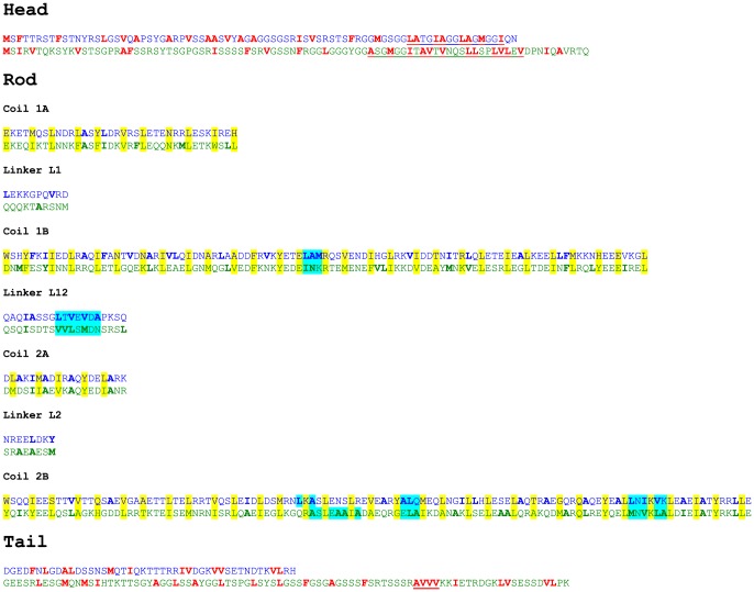
Figure 8. Amino acid sequence alignment of keratin assembly partners K18 (blue letters) and K8 (green letters).
The order of the subdomains is as reported in Figure 1 of [46]. Hydrophobic amino acids in the non-α-helical head and tail domains are indicated in red [47]. Significant hydrophobic motifs in these domains are underlined. In the rod domain, the a- and d-heptad positions are highlighted in yellow; these amino acids are responsible for the formation of a coiled-coil dimer from two individual α-helices. Hydrophilic domains on the surface of a coiled-coil dimer, generated by amino acids positioned in the b-, c-, e-, f-, and g-positions of the heptad pattern, are highlighted in cyan.

