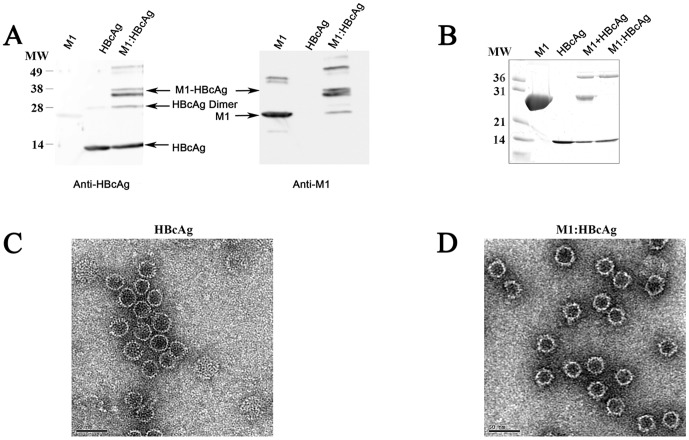
Figure 1. Synthesis and purification of M1:HBcAg VLPs.
M1 was coupled to HBcAg (residues 1–142) using the heterobifunctional cross-linker sMBS (Pierce). Western Blot indicating the coupled product is recognised by both the anti-M1 and anti-HBcAg antibodies. (B) Following conjugation, the M1:HBcAg was purified from unbound M1 protein by ultracentrifugation. The resulting coupled product was analysed by SDS-PAGE under reducing conditions followed by silver stain. Following purification no contaminating M1 was observed in the purified product. HBcAg (C) and M1:HBcAg (D) were stained with uranyl acetate and analysed by electron microscopy. The morphology of HBcAg was retained following coupling and purification.

