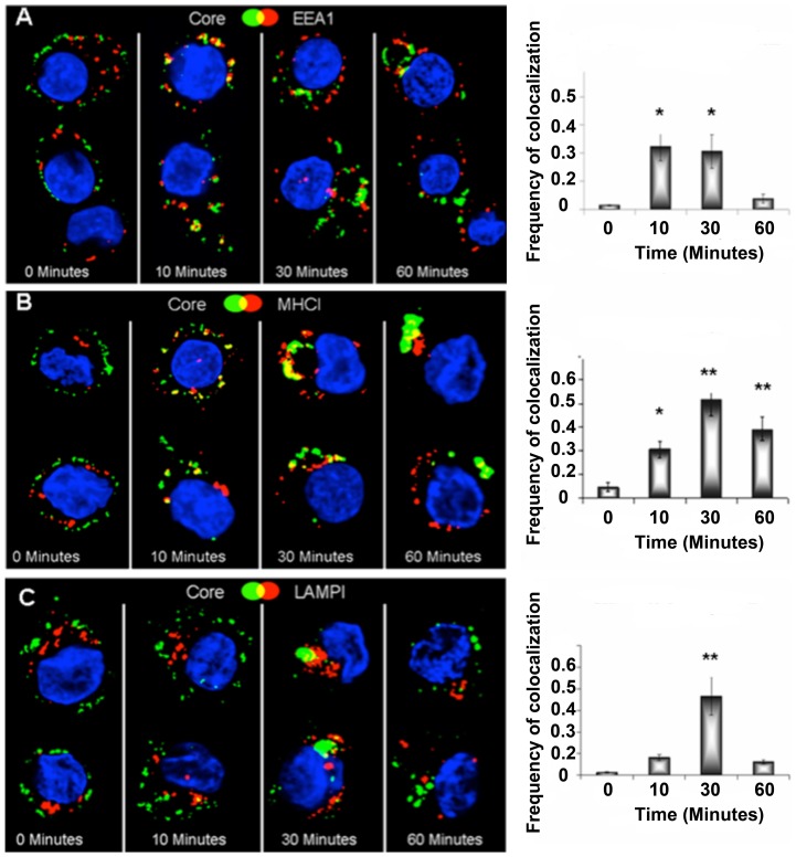
Figure 3. Colocalisation of M1:HBcAg and Endocytic markers.
Day 6 immature DCs were incubated with M1:HBcAg on ice to allow binding and the HBcAg was fluorescently labelled. The cells were subsequently incubated at 37°C to allow internalisation. At the indicated timepoints, cells were harvested, fixed, permeabilised and stained for endocytic markers (in green) for analysis by confocal microscopy. M1:HBcAg colocalised with the early endosome marker EEA1 within 10 minutes and the late endosomal marker LAMP1 from 30 minutes. Following internalisation, M1:HBcAg was shown to colocalise with MHC I. The frequency of pixel colocalisation (on the left) was determined by Manders coefficient from 4–8 cells per treatment. Statistical significance was determined by the unpaired student t-test; *p<0.05, **p<0.005.

