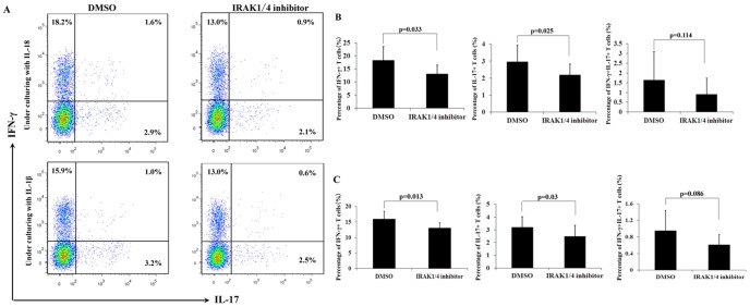
Figure 4. The effect of IRAK1/4 inhibitor on the expansion of Th1 cells and Th17 cells.
Purified CD4+T cells from normal controls (n = 8) were cultured with or without IRAK1/4 inhibitor for 3 days in the presence of wither IL-18 or IL-1β. The frequency of Th1 and Th17 cells was analyzed by FCM. A. A representative patient with data near the mean of each group in B and C. B. The results represent the percentages of IFN-γ+, IL-17+, and IL-17+IFN-γ+ cells among the CD4+T cells in the presence of IL-18. Results are expressed as means ± SD. C. The results represent the percentages of IFN-γ+,IL-17+, and IL-17+IFN-γ+ cells among the CD4+T cells in the presence of IL-1β. Results are expressed as means ± SD.

