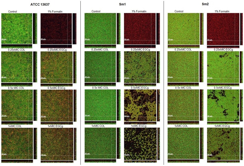
Figure 6. Optical sections of 48-h-old Sm biofilms (reference strain ATCC13637 and clinical isolates: Sm1 and Sm2) treated with EGCg and COL at 0.25×MIC, 0.5×MIC, 1×MIC.
Biofilms were treated with formalin as killing control. Live bacteria are stained in green (Syto9), dead bacteria in red (propidium iodide [PI]) or yellow (overlapping regions). Experiments were performed in duplicates (image data: 1024 ×1024 pixel with a pixel-size of 0.284 μm; z-step-size: 2 μm). Length of size bar: 50 μm.

