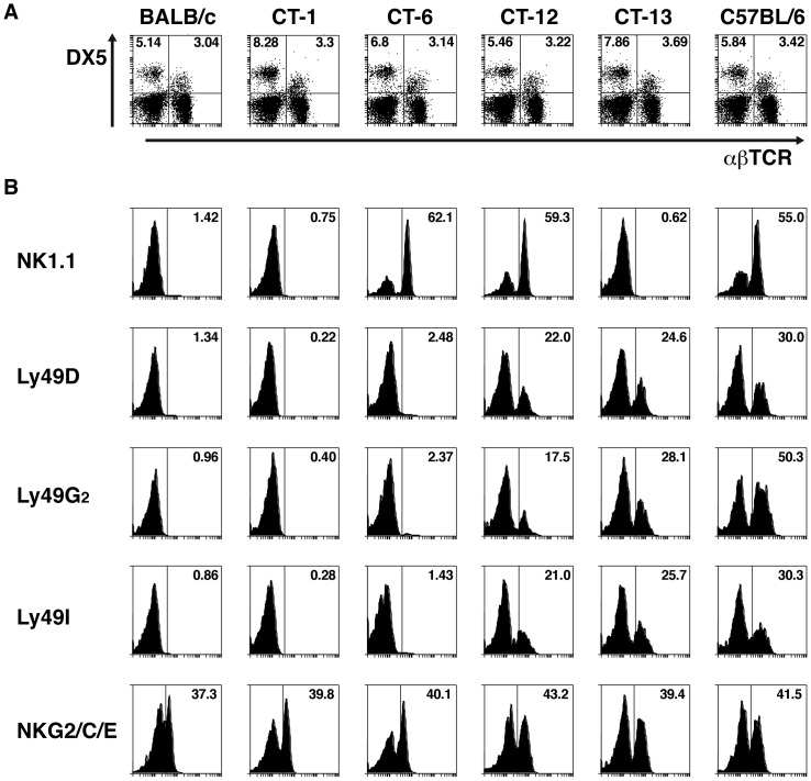
Figure 1. Differential expression of NKC markers in NK/NKT cells from BALB.B6-CT-1, BALB.B6-CT-6, BALB.B6-CT-12 and BALB.B6-CT-13 mice.
(A) Spleen cells from C57BL6, BALB/c and BALB.B6-CT-1, BALB.B6-CT-6, BALB.B6-CT-12 and BALB.B6-CT-13 mice were stained with anti-CD49b (DX5) and anti-αβ TCR antibodies. The percentage of NK and NKT cells are indicated. (B) The expression of the NKC markers NK1.1, Ly49A, Ly49D, Ly49G2, Ly49I, and NKG2A/C/E was calculated on all DX5 positive cells from the 6 mouse strains. Representative histograms are shown.

